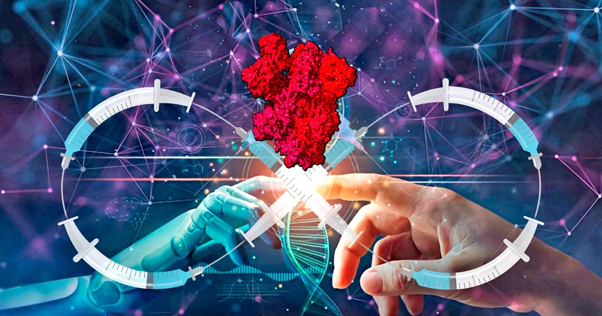
Las proteínas de la espícula (pico o spike), que originalmente forman parte de la capa externa del virus SARS-CoV2 y funcionan como una “llave” para “desbloquear” (infectar) las células, también son producidas en grandes cantidades por las “vacunas” de ARNm, lo que desencadena una respuesta inmunitaria de corta duración en forma de anticuerpos. Cada vez hay más pruebas de que la proteína de la espícula es dañina por sí misma (véase: 250 estudios sobre la patogenicidad de la proteína de la espícula, click aqui. Además, las investigaciones han demostrado que:
1) Tanto el ARNm de la “vacuna” que codifica el antígeno de la proteína pico o spike, como la proteína de pico en sí pueden penetrar tejidos distantes y causar daños sistémicos.
2) El ARNm de la “vacuna” y el antígeno de la proteína pico persisten en los tejidos de los receptores de vacunas humanas y de los sujetos de prueba animales mucho más tiempo de lo que afirman los funcionarios de salud pública, mientras que se ha demostrado que las proteínas pico virales persisten incluso más tiempo.
3) Las nanopartículas lipídicas ionizables (LNP) utilizadas en las inyecciones experimentales de ARNm son altamente inflamatorias por sí mismas, incluido su componente de polietilenglicol (PEG), una causa establecida de anafilaxia (una reacción alérgica extrema).
La siguiente colección de investigaciones presenta más de 130 estudios revisados por pares que documentan
- I) la amplia distribución y
- II) la persistencia del ARNm de la “vacuna” y la proteína de pico codificada, así como
- III) los posibles daños del sistema de administración de LNP (algunos estudios con hallazgos superpuestos aparecen en más de una categoría).
Tomados en conjunto con la evidencia de la patogenicidad de la proteína pico estos hallazgos sugieren que las “vacunas” de ARNm pueden distribuir la proteína pico dañina y de larga duración de manera incontrolable por todo el cuerpo, causando lesiones y muerte por diversos medios.
Tenga en cuenta que una pequeña cantidad de estudios en la sección:
- I) investigan la capacidad de la proteína pico viral resultante de la infección para cruzar barreras fisiológicas importantes por sí sola, mientras que algunos estudios en la sección
- II) demuestran la larga persistencia de la proteína de pico derivada del virus en ausencia de virus viable, lo que refuerza las preocupaciones sobre la proteína pico de la «vacuna» por ser similar.
Referencias
1. Australian Government Department of Health—Therapeutic Goods Administration, «Nonclinical
evaluation of BNT162b2 [mRNA] COVID-19 vaccine (COMIRNATY),» 2021, Available from: https://www.tga.gov.au/sites/default/files/foi-2389-06.pdf
• «Lipid nanoparticle formulation… and encapsulation efficiency similar to LNP in BNT162b2 /> vaccine… distribution mainly into liver, adrenal glands, spleen and ovaries over 48 h.»
2. Bansal S et al., «Cutting Edge: Circulating Exosomes with COVID Spike Protein Are Induced by /> BNT162b2 (Pfizer-BioNTech) Vaccination prior to Development of Antibodies: A Novel Mechanism /> for Immune Activation by mRNA Vaccines,» J. Immunol. 2021, 207, 10: 2405–2410. doi: /> https://doi.org/10.4049/jimmunol.2100637
• plasma
3. Baumeier C et al., «Intramyocardial Inflammation after COVID-19 Vaccination: An Endomyocardial /> Biopsy-Proven Case Series,» Int. J. Mol. Sci. 2022, 23, 13: 6940.
doi: https://doi.org/10.3390/ijms23136940
• «The expression of SARS-CoV-2 spike protein within the heart and the dominance of /> CD4+ lymphocytic infiltrates indicate an autoimmunological response to the vaccination.»
4. Boros LG et al., «Long-lasting, biochemically modified mRNA, and its frameshifted recombinant spike proteins in human tissues and circulation after COVID-19 vaccination,» Pharmacol Res Perspect 2024, 12, 3: e1218. doi: https://doi.org/10.1002/prp2.1218
• «… clinical studies now report that modified SARS-CoV-2 mRNA routinely persist up to a month from injection and can be detected in cardiac and skeletal muscle at sites of inflammation and fibrosis, while the recombinant spike protein may persist a little over half a year in blood.»
5. Brady M et al., «Spike protein multiorgan tropism suppressed by antibodies targeting SARS-CoV-2,»
Comm. Biol. 2021, 4, 1318. doi: https://doi.org/10.1038/s42003-021-02856-x
• After intravenous injection, «SP had a body-wide biodistribution, slow regional elimination, except for the liver, which showed an accumulation, and differential organ uptake. SP uptake was highest for the lungs and this was followed by the kidney, heart, and liver, but lowest in the brain.»
6. Brogna C et al., «Detection of recombinant Spike protein in the blood of individuals vaccinated
against SARS-CoV-2: Possible molecular mechanisms,» Proteonomics Clin App. 2023, 17, 6. doi:
https://doi.org/10.1002/prca.202300048
• plasma
7. Broudic K et al., «Nonclinical safety assessment of an mRNA Covid-19 vaccine candidate following repeated administrations and biodistribution,» J. Appl. Toxicol. 2024, 44, 3: 371-390. doi: https://doi.org/10.1002/jat.4548
• lymph nodes, spleen, liver, lacrimal glands, brain, thymus, lungs, adrenal glands, bone marrow, kidneys, testes, ovaries
8. Burkhardt A, «Pathology Conference: Vaccine-Induced Spike Protein Production in the Brain, Organs etc., now Proven.» Report24.news, 2022, available online: https://report24.news/pathologie-
konferenz-impfinduzierte-spike-produktion-in-gehirn-u-a-organen-nun-erwiesen/
• Heart, brain, liver, appendix, bronchi, skin, spleen
9. Buzhdygan TP et al., «The SARS-CoV-2 spike protein alters barrier function in 2D static and 3D microfluidic in-vitro models of the human blood–brain barrier,» Neurobiol Dis. 2020m 146: 105131.
doi: https://doi.org/10.1016/j.nbd.2020.105131
10. Castruita JAS et al., «SARS-CoV-2 spike mRNA vaccine sequences circulate in blood up to 28 days after COVID-19 vaccination,» APMIS 2023, 131: 128–132. doi: https://doi.org/10.1111/apm.13294
• plasma
11. Cosentino M and Franca Marino, «Understanding the Pharmacology of COVID- 19 mRNA Vaccines: Playing Dice with the Spike?» Int. J. Mol. Sci. 2022, 23, 18: 10881.
doi: https://doi.org/10.3390/ijms231810881
• «Taken as a whole, evidence strongly supports the possible link between inappropriate expression of S protein in sensitive tissues and subsequent tissue damage.»
12. DeOre BJ et al., «SARS-CoV-2 Spike Protein Disrupts Blood-Brain Barrier Integrity via RhoA Activation,» J Neuroimmune Pharmacol. 2021, 16, 4:722-728. Doi: https://doi.org/10.1007/s11481- 021-10029-0
13. Di J et al., «Biodistribution and Non-linear Gene Expression of mRNA LNPs Affected by Delivery Route and Particle Size,» Pharm Res 2022, 39: 105-114. doi: https://doi.org/10.1007/s11095-022-03166-5
• liver, spleen, muscle, and inguinal lymph nodes
14. European Medicines Agency, Assessment Report, available online: https://www.ema.europa.eu/en/documents/assessment-report/comirnaty-epar-public- assessment-report_en.pdf
• «Synthetic mRNAs encapsulated in LNPs can reach many organs, such as the spleen, heart, kidneys, lungs and brain. The mRNAs were found in the ovaries and the testicles in small quantities, during the biodistribution studies of this vaccine after 9 days.»
15. European Medicines Agency, COVID-19 Vaccine Moderna, available online: https://www.ema.europa.eu/en/documents/assessment-report/spikevax-previously-covid- 19-vaccine-moderna-epar-public-assessment-report_en.pdf
• Vaccine mRNAs are detectable in a wide variety of organs: brain, heart, lungs, eyes, gonads.
16. Fertig TE et al., «Beyond the injection site: identifying the cellular targets of mRNA vaccines,» J Cell Ident 2024, 3, 1. doi: 10.47570/joci.2024.004
• Overview of studies showing wide distribution throughout the body.
17. Fertig TE et al., «Vaccine mRNA Can Be Detected in Blood at 15 Days Post Vaccination,» Biomedicines 2022, 10, 7: 1538. doi: https://doi.org/10.3390/biomedicines10071538
• plasma
18. Hanna N et al. «Biodistribution of mRNA COVID-19 vaccines in human breast milk,» eBioMedicine 2023, 96, 104800. doi: 10.1016/j.ebiom.2023.104800
• «Of 13 lactating women receiving the vaccine (20 exposures), trace mRNA amounts were detected in 10 exposures up to 45 h post-vaccination. «
19. Hano S et al., «A case of persistent, confluent maculopapular erythema following a COVID-19 mRNA vaccination is possibly associated with the intralesional spike protein expressed by vascular endothelial cells and eccrine glands in the deep dermis,» J Dermatol 2023, 50, 9: 1208-1212. doi:
https://doi.org/10.1111/1346-8138.16816
• «Surprisingly, immunohistochemical staining of the lesion 100 days after the disease onset revealed the COVID-19 spike protein expressed by vascular endothelial cells and eccrine glands in the deep dermis. As she had no episode of COVID-19 infection, it is highly likely that the spike protein was derived from the mRNA vaccine and it might be the cause of the development and persistence of her skin lesions.»
20. Hulscher N et al., «Autopsy findings in cases of fatal COVID-19 vaccine-induced myocarditis,» ESC Heart Failure 2024. doi: https://doi.org/10.1002/ehf2.14680
• «COVID-19 vaccine Spike protein is produced in the body for an uncontrolled duration and in
unknown quantity resulting in deleterious effects, especially on the heart, explaining the cardiovascular deaths seen in our study without evidence of other organ system involvement.»
21. Judicial Watch, «JW v HHS FDA Pfizer BioNTech Vaccine prod 3 02418,» March 21, 2022,
https://www.judicialwatch.org/documents/jw-v-hhs-fda-pfizer-biontech-vaccine-prod-3-02418/
• LNP biodistribution to liver, spleen, adrenal glands, ovaries. «Outside the injection site, low levels of radioactivity were detected in most tissues, with the greatest levels in plasma observed 1-4 hours post-dose.»
22. Kammala AK et al., «In vitro mRNA-S maternal vaccination induced altered immune regulation at the
maternal-fetal interface,» Am. J. Reprod. Immunol. 2024, 91, 5: e13861.
doi: https://doi.org/10.1111/aji.13861
• «… our study indicates that mRNA-S-based maternal vaccination during pregnancy may
influence the maternal-fetal interface’s COVID-19 interaction and immune regulation. Further
investigation is warranted to assess safety and implications.»
23. Kawano H et al., «Fulminant Myocarditis 24 Days after Coronavirus Disease Messenger Ribonucleic Acid Vaccination,» Intern. Med. 2022, 61, 15: 2319-2325. doi:
https://doi.org/10.2169/internalmedicine.9800-22
• «… positive immunostaining for severe acute respiratory syndrome coronavirus 2 spike protein and C4d in the myocardium.»
24. Kent SJ et al., «Blood Distribution of SARS-CoV-2 Lipid Nanoparticle mRNA Vaccine in Humans,» ACS Nano 2024, 18, 39: 27077-27089. doi: https://doi.org/10.1021/acsnano.4c11652
• «The similar kinetics of intact mRNA and the ionizable lipid in blood and the slow degradation of the mRNA suggest that mRNA lipid nanoparticles remain intact and travel from injection sites or lymph nodes into the bloodstream within 4 h postvaccination. The rapid dissemination of mRNA lipid nanoparticles in blood found in our study is consistent with the recent findings on the detection of mRNA in breast milk at 3−45 h postvaccination.»
25. Krauson AM et al., «Duration of SARS-CoV-2 mRNA vaccine persistence and factors associated with
cardiac involvement in recently vaccinated patients,» npj Vaccines, 8, 141. doi:
https://doi.org/10.1038/s41541-023-00742-7
• axillary lymph nodes, myocardium
26. Kwon MH et al., «The Pharmacokinetics of mRNA Vaccine Carrier using Carbon-14,» J. Radiopharm. Mol. Probes 2024, 10, 1: 73-81. doi: https://doi.org/10.22643/JRMP.2024.10.1.73
• serum, lymph nodes, muscle, spleen, liver, testis, ovary, thymus, lung, brain
27. Lehmann KJ, «SARS-CoV-2-Spike Interactions with the Renin-Angiotensin-Aldosterone System – Consequences of Adverse Reactions of Vaccination,» J Biol Today’s World 2023, 12/4: 001- 013. https://doi.org/10.31219/osf.io/27g5h
• «The presented analysis provides a substantial body of evidence for the causal involvement of Ang II/activated RAAS in eliciting adverse reactions after application of spike-inducing vaccine.
As an example, some serious organ disturbances or adverse reactions, in which the connection with an activated RAAS is obvious (cardiovascular and blood coagulation disorders, disorders of the nervous and muscular system, inflammatory reactions, auto-immunological, vascular and renal disorders), are presented and discussed…»
28. Li C. et al., «Intravenous Injection of Coronavirus Disease 2019 (COVID-19) MRNA Vaccine Can Induce Acute Myopericarditis in Mouse Model,» Clin. Infect. Dis. 2022, 74, 11: 1933-1950. doi:
https://doi.org/10.1093/cid/ciab707
• «Severe acute respiratory syndrome coronavirus 2 (SARS-CoV-2) spike antigen expression by immunostaining was occasionally found in infiltrating immune cells of the heart or injection site, in cardiomyocytes and intracardiac vascular endothelial cells, but not skeletal myocytes.»
29. Li C et al., «Mechanisms of innate and adaptive immunity to the Pfizer-BioNTech BNT162b2
vaccine,» Nature Immunol. 2022, 23: 543-555. doi: https://doi.org/10.1038/s41590-022-01163-9 • spleen, muscle, liver, lung and non-dLNs
30. Lin X et al., «Transplacental transmission of the COVID-19 vaccine messenger RNA: evidence from
placental, maternal, and cord blood analyses postvaccination,» Am J Obstet Gynecol 2024, 92, 4:
e13934. doi: https://doi.org/10.1111/aji.13934
• «The vaccine mRNA was detected in the 2 placentas evaluated using quantitative ddPCR and
ISH… Using WES, the spike protein expression was detected in the placenta of patient 2, but not
in patient 1… Furthermore, the vaccine mRNA was detected in the umbilical cord and maternal
blood of patient 1 using ddPCR.»
31. Luo Y et al., «SARS-Cov-2 spike induces intestinal barrier dysfunction through the interaction between CEACAM5 and Galectin-9,» Front. Immunol. 2024, 15. doi:
https://doi.org/10.3389/fimmu.2024.1303356
32. Magen E et al., «Clinical and Molecular Characterization of a Rare Case of BNT162b2 mRNA COVID- 19 Vaccine-Associated Myositis,» Vaccines 2022, 10, 7: 1135.
doi: https://doi.org/10.3390/vaccines10071135
• «… although the BNT162b2 vaccine mRNA was not properly expressed in blood cells seven days after receipt of the first vaccine dose, it was still expressed in muscle tissue distant from the vaccination site one month after receipt of the first vaccine dose.»
33. Magro C et al., «The histologic and molecular correlates of COVID-19 vaccine-induced changes in the skin,» Clin. Dermatol. 2021, 39, 6: 966-984. doi:
https://doi.org/10.1016/j.clindermatol.2021.07.011
• Spike detected in deep dermis or blood vessels serving skin in 10/34 cases.
34. Magro C et al., «Disruption of the blood-brain barrier is correlated with spike endocytosis by ACE2 + endothelia in the CNS microvasculature in fatal COVID-19. Scientific commentary on ‘Detection of blood-brain barrier disruption in brains of patients with COVID-19, but no evidence of brain penetration by SARS-CoV-2’,» Acta Neuropathol. 2024, 147, 1: 47. doi:
https://doi.org/10.1007/s00401-023-02681-y
35. Martin-Navarro L et al., «In situ detection of vaccine mRNA in the cytoplasm of hepatocytes during COVID-19 vaccine-related hepatitis,» J. Hepatol. 2023, 78, 1: e20-e22.
doi: 10.1016/j.jhep.2022.08.039
• «… our results suggest that lipid nanoparticles bearing mRNA molecules encoding SARS-CoV-2 proteins can reach hepatocytes under certain circumstances and deliver mRNA in high quantities that could be used by the translational machinery of the cells to produce spike.»
36. Maugeri M et al.. «Linkage between endosomal escape of LNP-mRNA and loading into EVs for transport to other cells,» Nat Commun 2019, 10: 4333. doi: https://doi.org/10.1038/s41467-019- 12275-6
• «The present study shows that LNP components (mRNA and ionizable lipids) are partly incorporated into endo-EVs… these endo-EVs protect exogenous mRNA during in vivo transport to organs…»
37. Medicines & Healthcare Products Regulatory Agency, «Summary of the Public Assessment Report for Pfizer/BioNTech COVID-19 Vaccine,» available online: https://www.gov.uk/government/publications/regulatory-approval-of-pfizer-biontech- vaccine-for-covid-19/summary-public-assessment-report-for-pfizerbiontech-covid-19-vaccine
• liver
38. Ministry of Health, Labour and Welfare of Japan, «SARS-CoV-2 mRNA Vaccine (BNT162, PF- 07302048): Summary Text of the Pharmacokinetic Study,» available online:
https://www.docdroid.net/xq0Z8B0/pfizer-report-japanese-government-pdf
• bladder, bone, bone marrow, brain, eyes, heart, kidneys, large intestine, liver, lung
39. Mörz M, «A Case Report: Multifocal Necrotizing Encephalitis and Myocarditis after BNT162b2 mRNA Vaccination against COVID-19,» Vaccines 2022, 10, 10: 1651.
doi: https://doi.org/10.3390/vaccines10101651
• «Only spike protein but no nucleocapsid protein could be detected within the foci of inflammation in both the brain and the heart, particularly in the endothelial cells of small blood vessels.»
40. Nyein CM et al., «Severe de novo liver injury after Moderna vaccination – not always autoimmune hepatitis,» J. Hepatol. 2022, 77, 2: 556-558. Doi: 10.1016/j.jhep.2022.03.041
• «Unique to this case is the demonstration of anti-SARS-CoV-2 spike protein within the liver parenchyma.»
41. Ogata AF et al., «Circulating Severe Acute Respiratory Syndrome Coronavirus 2 (SARS-CoV-2) Vaccine Antigen Detected in the Plasma of mRNA-1273 Vaccine Recipients,» Clin. Infect. Dis. 2022, 75, 4: 715–718. doi: https://doi.org/10.1093/cid/ciab465
• «Here we provide evidence that circulating SARS-CoV-2 proteins are present in the plasma of participants vaccinated with the mRNA-1273 vaccine.»
42. Pateev I et al., «Biodistribution of RNA Vaccines and of Their Products: Evidence from Human and Animal Studies,» Biomedicines 2024, 12, 1: 59. doi: https://doi.org/10.3390/biomedicines12010059
• «Intravenous injection led to the detection of fluorescent proteins in the liver, spleen, lungs, and lymph nodes.»
43. Petrovszki D et al., «Penetration of the SARS-CoV-2 Spike Protein across the Blood-Brain Barrier, as Revealed by a Combination of a Human Cell Culture Model System and Optical Biosensing,» Biomedicines 2022, 10, 1: 188. doi: https://doi.org/10.3390/biomedicines10010188
44. Rhea EM et al., «The S1 protein of SARS-CoV-2 crosses the blood–brain barrier in mice,» Nature Neuroscience 2021, 24, 3: 368–378. doi: https://doi.org/10.1038/s41593-020-00771-8
45. Röltgen K et al., «Immune imprinting, breadth of variant recognition, and germinal center response in human SARS-CoV-2 infection and vaccination,» Cell, 2022, 185, 6: 1025-1040. doi: 10.1016/j.cell.2022.01.018
• «mRNA vaccination stimulates robust GCs containing vaccine mRNA and spike antigen up to 8 weeks postvaccination in some cases.»
46. Rzymski P and Andrzej Fal, «To aspirate or not to aspirate? Considerations for the COVID-19 vaccines,» Pharmacol. Rep 2022, 74: 1223–1227. doi: https://doi.org/10.1007/s43440-022-00361-4
• «As shown in vivo in mice, intravenous injection of the BNT162b2 vaccine (BioNTech/Pfizer, Germany/USA) resulted in histopathological changes characteristic for myopericarditis… the amount of mRNA encoding SARS-CoV-2 spike protein and its subsequent myocardial expression was significantly higher in heart tissue when compared to the animals receiving the intramuscular injection.»
47. Sandelius A et al., «Biodistribution of lipid nanoparticle, eGFP mRNA and translated protein following subcutaneous administration in mouse,» Bioanalysis 2024, 16, 14: 721-733. doi:
https://doi.org/10.1080/17576180.2024.2360361
• skin, spleen, liver, kidney
48. Sano H et al., «A case of persistent, confluent maculopapular erythema following a COVID-19 mRNA vaccination is possibly associated with the intralesional spike protein expressed by vascular endothelial cells and eccrine glands in the deep dermis,» J Dermatol 2023, 50, 9: 1208-1212. doi:
https://doi.org/10.1111/1346-8138.16816
• «Surprisingly, immunohistochemical staining of the lesion 100 days after the disease onset revealed the COVID-19 spike protein expressed by vascular endothelial cells and eccrine glands in the deep dermis. As she had no episode of COVID-19 infection, it is highly likely that the spike protein was derived from the mRNA vaccine and it might be the cause of the development and persistence of her skin lesions.»
49. Sano S et al., «SARS-CoV-2 spike protein found in the acrosyringium and eccrine gland of repetitive miliaria-like lesions in a woman following mRNA vaccination,» J. Dermatol. 2024, 51, 9: e293-e295.
doi: https://doi.org/10.1111/1346-8138.17204
• cutaneous
50. Sattar S Et al., «Nuclear translocation of spike mRNA and protein is a novel feature of SARS-CoV-2,» 2023 Front. Microbiol. 2023, 14 (Sec. Virology). doi: https://doi.org/10.3389/fmicb.2023.1073789
• «Although the S protein is a surface transmembrane type 1 glycoprotein, it has been predicted to be translocated into the nucleus due to the novel nuclear localization signal (NLS) «PRRARSV,» which is absent from the S protein of other coronaviruses. Indeed, S proteins translocate into the nucleus in SARS-CoV-2-infected cells. S mRNAs also translocate into the nucleus. S mRNA colocalizes with S protein, aiding the nuclear translocation of S mRNA.»
51. Schreckenberg R et al., «Cardiac side effects of RNA-based SARS-CoV-2 vaccines: Hidden cardiotoxic effects of mRNA-1273 and BNT162b2 on ventricular myocyte function and structure,» Br. J. Pharmacol. 2024, 181, 3: 345-361. doi: https://doi.org/10.1111/bph.16262
• «After 48 h, expression of the encoded spike protein was detected in ventricular cardiomyocytes for both mRNAs. At this point in time, mRNA-1273 induced arrhythmic as well as completely irregular contractions associated with irregular as well as localized calcium transients, which provide indications of significant dysfunction of the cardiac ryanodine receptor (RyR2). In contrast, BNT162b2 increased cardiomyocyte contraction via significantly increased protein kinase A (PKA) activity at the cellular level.»
52. Stern B et al., «SARS-CoV-2 spike protein induces endothelial dysfunc?on in 3D engineered vascular networks.» J. Biomed. Mater. Res. A. 2023, 112, 4: 524-533. doi: hJps://doi.org/10.1002/jbm.a.37543
53. Suprewicz L et al., «Blood-brain barrier function in response to SARS-CoV-2 and its spike protein,» Neurol. Neurochir Pol. 2023, 57: 14–25. doi: 10.5603/PJNNS.a2023.0014
• «…the S-protein may interact directly with the BBB, and S1, S1RBD, and S2 subunits exhibit pro- inflammatory effects, resulting in increased BBB permeability via damage to tight junctions (TJs) but not adherens junctions (AJs).»
54. Suprewicz L et al., «Recombinant human plasma gelsolin reverses increased permeability of the blood- brain barrier induced by the spike protein of the SARS-CoV-2 virus,» J Neuroinflamma5on 2022, 19, 1: 282,
doi: hJps://doi.org/10.1186/s12974-022-02642-4
55. Takanashi A et al., «Delivery and Expression of mRNA in the Secondary Lymphoid Organs Drive Immune Responses to Lipid Nanoparticle-mRNA Vaccines after Intramuscular Injection,» Mol. Pharmaceutics 2023, 20, 8: 3876–3885. doi: https://doi.org/10.1021/acs.molpharmaceut.2c01024
• «Our results suggest that the mRNA delivery and transfection of secondary lymphatic organs, not LNP adjuvancy or RNA expression in muscle, are the main drivers for adaptive immune response in mice.»
56. Yamamoto M et al., «Persistent varicella zoster virus infection following mRNA COVID-19 vaccination was associated with the presence of encoded spike protein in the lesion,» J. Cutan.
Immunol. Allergy 2022, 6, 1: 18-23. doi: https://doi.org/10.1002/cia2.12278
• «Strikingly, the vaccine-encoded spike protein of the COVID-19 virus was expressed in the vesicular keratinocytes and endothelial cells in the dermis.»
57. Yonker LM et al., «Circulating Spike Protein Detected in Post–COVID-19 mRNA Vaccine Myocarditis,» Circulation 2023, 147, 11. doi: https://doi.org/10.1161/CIRCULATIONAHA.122.061025
• plasma
II. Spike protein and vaccine mRNA persistence research library
Compiled by Dr. Martin Wucher, MSC Dent Sc (eq DDS), Erik Sass, et al.
Dozens of studies collected here (n=39) demonstrate that both «vaccine» mRNA, and the spike protein antigen it encodes, persist in the tissues of human vaccine recipients and animal test subjects far longer than claimed by public health officials: up to eight weeks in the case of mRNA (Röltgen K et al.) and up to six months for spike protein (Brogna C et al.).
Numerous studies have also shown that viral spike proteins can persist even longer in individuals recovered from SARS CoV2 infection or with «long COVID,» with spike protein detected 15 months (Patterson BK et al.) to two years (Fraser ME at al.) after infection.
Long-lasting viral spike proteins have frequently been detected in the absence of viable virus, as reflected in negative PCR tests and RNA assays, suggesting identical «vaccine» spike proteins may also persist for a year or more.
This compilation originated with Dr. Wucher’s contribution to TOXIC SHOT: Facing the Dangers of the COVID «Vaccines,» (Chapter 4: The Spike Protein Is Harmful By Itself).
ANNOTATED REFERENCES (n=39)
1. Bansal S, et al. «Cutting Edge: Circulating Exosomes with COVID Spike Protein Are Induced by BNT162b2 (Pfizer-BioNTech) Vaccination prior to Development of Antibodies: A Novel Mechanism for Immune Activation by mRNA Vaccines,» J. Immunol. 2021, 207, 10: 2405–2410. doi:
https://doi.org/10.4049/jimmunol.2100637
• circulating exosomes with spike protein detected four months after vaccination.
2. Boros LG et al., «Long-lasting, biochemically modified mRNA, and its frameshifted recombinant spike proteins in human tissues and circulation after COVID-19 vaccination,» Pharmacol Res Perspect 2024, 12, 3: e1218. doi: https://doi.org/10.1002/prp2.1218
• «… clinical studies now report that modified SARS-CoV-2 mRNA routinely persist up to a month from injection and can be detected in cardiac and skeletal muscle at sites of inflammation and fibrosis, while the recombinant spike protein may persist a little over half a year in blood.»
3. Brogna C et al., «Detection of recombinant Spike protein in the blood of individuals vaccinated against SARS-CoV-2: Possible molecular mechanisms,» Proteonomics Clin App. 2023, 17, 6. doi:
https://doi.org/10.1002/prca.202300048
• «The minimum and maximum time at which PP-Spike was detected after vaccination was 69 and 187 days, respectively.»
4. Castruita JAS et al., «SARS-CoV-2 spike mRNA vaccine sequences circulate in blood up to 28 days after COVID-19 vaccination,» APMIS 2023, 131: 128–132. doi: https://doi.org/10.1111/apm.13294
5. Cheung CCL et al., «Residual SARS-CoV-2 viral antigens detected in GI and hepatic tissues from five recovered patients with COVID-19,» Gut 2022, 71, 1: 226–9. doi: https://doi.org/10.1136/gutjnl-2021- 324280
• Persistence of residual SARS-CoV-2 antigens up to 180 days in the colon, appendix, ileum, haemorrhoid, liver, gallbladder and lymph nodes; unable to detect viral RNA in many patients’ tissues.
6. Colmenero I et al., «SARS-CoV-2 endothelial infection causes COVID-19 chilblains: histopathological, immunohistochemical and ultrastructural study of seven paediatric cases,» Br J Dermatol. 2020, 183: 729-737. doi: https://doi.org/10.1111/bjd.19327
• Spike protein detected in lesions up to 30 days after onset of acute infection. SARS-CoV-2 PCR from nasopharyngeal and oropharyngeal swabs was negative in all cases tested (six of six).
7. Craddock V et al., «Persistent circulation of soluble and extracellular vesicle-linked Spike protein in individuals with postacute sequelae of COVID-19,» J Med. Virol. 2023, 95, 2: e28568. doi:
https://doi.org/10.1002/jmv.28568
• «… our findings suggest that Spike and/or viral RNA fragments persist in the recovered COVID-19 patients with PASC up to 1 year or longer after acute SARS-CoV-2 infection.» Further, «this is the first report to show that part of the circulating Spike is linked to extracellular vesicles without any presence of viral RNA in these vesicles.»
8. European Medicines Agency, Assessment Report, available online: https://www.ema.europa.eu/en/documents/assessment-report/comirnaty-epar-public- assessment-report_en.pdf
• «Synthetic mRNAs encapsulated in LNPs can reach many organs, such as the spleen, heart, kidneys, lungs and brain. The mRNAs were found in the ovaries and the testicles in small quantities, during the biodistribution studies of this vaccine after 9 days…»
9. Fertig TE et al., «Vaccine mRNA Can Be Detected in Blood at 15 Days Post Vaccination,» Biomedicines 2022, 10, 7: 1538. doi: https://doi.org/10.3390/biomedicines10071538
10. Fraser ME at al., «SARS-CoV-2 Spike Protein and Viral RNA Persist in the Lung of Patients With Post- COVID Lung Disease (abstract),» Am J Respir Crit Care Med 2024, 209: A4193. doi:
https://doi.org/10.1164/ajrccm-conference.2024.209.1_MeetingAbstracts.A4193
• «Spike protein and RNA persists in BAL from patients with post-COVID lung disease up to two years after acute infection.»
11. Gaebler C et al., «Evolution of antibody immunity to SARS-CoV-2,» Nature 2021, 591: 639-644. doi:
https://doi.org/10.1038/s41586-021-03207-w
• Gastrointestinal tract biopsies suggest spike antigen persisted in the small bowel in 7 of 14 individuals who were asymptomatic at 4 months after infection… Clinically approved nasopharyngeal-swab PCR assays were negative in all 14 individuals at the time of biopsy.
However, biopsy samples from 3 of the 14 participants produced PCR amplicons that were sequence-verified as SARS-CoV-2. In addition, viral RNA was detected by in situ hybridization in biopsy samples from the two participants who were tested.
12. George S et al., «Evidence for SARS-CoV-2 Spike Protein in the Urine of COVID-19 Patients,» Kidney360 2021, 2, 6: 924-936. doi: 10.34067/KID.0002172021
• «The SARS-CoV-2 spike protein could be detected in urine from day 1 to day 44 post–hospital admission… Of the 23 adults who were Ur-S+, only one individual showed detectable viral RNA in urine.»
13. Goh D et al., «Case report: Persistence of residual antigen and RNA of the SARS-CoV-2 virus in
tissues of two patients with long COVID,» Front. Immunol. 2022, 13 (Sec. Viral Immunology). doi:
https://doi.org/10.3389/fimmu.2022.939989
• Persistence of spike protein 426 days after symptom onset; residual viral RNA also detected.
14. Hano S et al., «A case of persistent, confluent maculopapular erythema following a COVID-19 mRNA vaccination is possibly associated with the intralesional spike protein expressed by vascular endothelial cells and eccrine glands in the deep dermis,» J Dermatol 2023, 50, 9: 1208-1212. doi:
https://doi.org/10.1111/1346-8138.16816
• «Surprisingly, immunohistochemical staining of the lesion 100 days after the disease onset revealed the COVID-19 spike protein expressed by vascular endothelial cells and eccrine glands in the deep dermis. As she had no episode of COVID-19 infection, it is highly likely that the spike protein was derived from the mRNA vaccine and it might be the cause of the development and persistence of her skin lesions.»
15. Karaba AH et al., «Detectable plasma severe acute respiratory syndrome coronavirus 2 spike antigen is associated with poor antibody response following third messenger RNA vaccination in kidney transplant recipients,» Transpl Infect Dis 2024, 26, 3: e14281. doi: https://doi.org/10.1111/tid.14281
• Spike protein detectable in 3/16 (19%) participants 14 days after vaccination.
16. Kawano H et al., «Fulminant Myocarditis 24 Days after Coronavirus Disease Messenger Ribonucleic Acid Vaccination,» Intern. Med. 2022, 61, 15: 2319-2325. doi:
https://doi.org/10.2169/internalmedicine.9800-22
• «… positive immunostaining for severe acute respiratory syndrome coronavirus 2 spike protein and C4d in the myocardium.»
17. Kent SJ et al., «Blood Distribution of SARS-CoV-2 Lipid Nanoparticle mRNA Vaccine in Humans,» ACS Nano 2024, 18, 39: 27077-27089. doi: https://doi.org/10.1021/acsnano.4c11652
• «The vaccine mRNA was detectable and quantifiable up to 14–15 days postvaccination in 37% of subjects.»
18. Krauson AM et al., «Duration of SARS-CoV-2 mRNA vaccine persistence and factors associated with cardiac involvement in recently vaccinated patients,» npj Vaccines, 8, 141. doi:
https://doi.org/10.1038/s41541-023-00742-7
• «Vaccine was detected in the axillary lymph nodes in the majority of patients dying within 30 days of vaccination… Vaccine was detected in the myocardium in a subset of patients vaccinated within 30 days of death.»
19. Li C et al., «Mechanisms of innate and adaptive immunity to the Pfizer-BioNTech BNT162b2 vaccine,» Nature Immunol. 2022, 23: 543-555. doi: https://doi.org/10.1038/s41590-022-01163-9
• «mRNA could be detected in the spleen, and the spike protein itself was detectable in the serum, for up to 7 d after immunization.»
20. Magen E et al., «Clinical and Molecular Characterization of a Rare Case of BNT162b2 mRNA COVID- 19 Vaccine-Associated Myositis,» Vaccines 2022, 10, 7: 1135.
doi: https://doi.org/10.3390/vaccines10071135
• «… although the BNT162b2 vaccine mRNA was not properly expressed in blood cells seven days after receipt of the first vaccine dose, it was still expressed in muscle tissue distant from the vaccination site one month after receipt of the first vaccine dose.»
21. Mayordomo-Colunga J et al., «SARS-CoV-2 spike protein in intestinal cells of a patient with
coronavirus disease 2019 multisystem inflammatory syndrome,» J Pediatr. 2022, 243: 214-18e215.
doi: https://doi.org/10.1016/j.jpeds.2021.11.058
• Spike protein detected 6 weeks after acute infection. «At presentation, the patient tested
negative for SARS-CoV-2 by reverse-transcriptase polymerase chain reaction on nasopharyngeal
swab but positive for serum SARS-CoV-2 immunoglobulin G.»
22. Mörz M, «A Case Report: Multifocal Necrotizing Encephalitis and Myocarditis after BNT162b2 mRNA Vaccination against COVID-19,» Vaccines 2022, 10, 10: 1651.
doi: https://doi.org/10.3390/vaccines10101651
• Vaccine-induced spike detected on autopsy three weeks after last injection.
23. Ogata AF et al., «Circulating Severe Acute Respiratory Syndrome Coronavirus 2 (SARS-CoV-2) Vaccine Antigen Detected in the Plasma of mRNA-1273 Vaccine Recipients,» Clin. Infect. Dis. 2022, 74, 4: 715-728. doi: https://doi.org/10.1093/cid/ciab465
• «Spike protein was detectable in 3 of 13 participants an average of 15 days after the first injection.»
24. Parcial ALN et al., «SARS-CoV-2 Is Persistent in Placenta and Causes Macroscopic, Histopathological, and Ultrastructural Changes,» Viruses 2022, 14, 9: 1885.
doi: https://doi.org/10.3390/v14091885
• «Three of five placentas presented SARS-CoV-2 RNA detected by RT-PCRq at least two to twenty weeks after primary pregnancy infection symptoms, and SARS-CoV-2 spike protein was detected in all placentas by immunoperoxidase assay.»
25. Pateev I et al., «Biodistribution of RNA Vaccines and of Their Products: Evidence from Human and Animal Studies,» Biomedicines 2024, 12, 1: 59. doi: https://doi.org/10.3390/biomedicines12010059
• (Roltgen K et al) «The amount of the spike antigen declined significantly at 4 months after the double vaccination but was still detectable.»
• «Immunohistochemical staining for the spike antigen in the lymph nodes of vaccinated patients revealed peak amounts of the spike protein in germinal centers 16 days after dose 2, with the spike antigen still detectable on day 60.»
• (Brogna C et al.) «It is noteworthy that in this study, spike protein was still detected in human
blood on the 187th day after vaccination.»
26. Patterson BK et al., «Persistence of SARS CoV-2 S1 Protein in CD16+ Monocytes in Post-Acute Sequelae of COVID-19 (PASC) up to 15 Months Post-Infection,» Front. Immunol. 2022, 12: 746021.
doi: https://doi.org/10.3389/fimmu.2021.746021
• Intact viral RNA undetectable in monocytes.
27. Peluso MJ et al., «Plasma-based antigen persistence in the post-acute phase of COVID-19,» Lancet 2024, 24, 6: E345-E347. doi: 10.1016/S1473-3099(24)00211-1
• «Of 660 pandemic-era specimens tested, 61 (9·2%) specimens from 42 participants (25% of the group), had one or more detectable SARS-CoV-2 antigens. The most commonly detected antigen was spike (n=33, 5·0%), followed by S1 (n=15, 2·3%)…»
• «… our data provide strong evidence that SARS-CoV-2, in some form or location, persists for up to 14 months following acute SARS-CoV-2 infection.» • «… our findings provide no direct evidence regarding the persistent presence of replication- competent or even transcriptionally active virus.»
28. Peluso MJ et al., «SARS-CoV-2 and mitochondrial proteins in neural-derived exosomes of COVID- 19,» Ann Neurol 2022, 91, 6: 772-781. doi: https://doi.org/10.1002/ana.26350
• Exosomes containing spike protein were detected in plasma of long COVID patients with neuropsychiatric symptoms at two months.
29. Roden AC et al., «Comparison of In Situ Hybridization, Immunohistochemistry, and Reverse Transcription–Droplet Digital Polymerase Chain Reaction for Severe Acute Respiratory Syndrome Coronavirus 2 (SARS-CoV-2) Testing in Tissue,» Arch Pathol Lab Med 2021, 145, 7: 785–796. doi:
https://doi.org/10.5858/arpa.2021-0008-SA
• Detected viral protein 46 days after onset of symptoms.
• «All patients from our institution had tested positive for COVID-19 by nasopharyngeal swab within a median of 14.5 days (range, 0–67 days) before death. All patients from our institution but one were tested for COVID-19 again at time of autopsy; 10 of 13 (76.9%) tested positive.»
30. Röltgen K et al., «Immune imprinting, breadth of variant recognition, and germinal center response in human SARS-CoV-2 infection and vaccination,» Cell, 2022, 185, 6: 1025-1040. doi:
10.1016/j.cell.2022.01.018
• «mRNA vaccination stimulates robust GCs containing vaccine mRNA and spike antigen up to 8 weeks postvaccination in some cases.»
• «… with spike antigen still present as late as 60 days post-second dose»
31. Rong Z et al., «Persistence of spike protein at the skull-meninges-brain axis may contribute to the neurological sequelae of COVID-19,» Cell Host Microbe 2024, 26: S1931-3128(24)00438-4. doi: 10.1016/j.chom.2024.11.007
• «In a time course experiment, we found the spike protein in the skull marrow, kidney, liver, and lung 3 days post-injection, remaining detectable in the kidney and liver 14 days post-injection.»
32. Sano H et al., «A case of persistent, confluent maculopapular erythema following a COVID-19 mRNA
vaccination is possibly associated with the intralesional spike protein expressed by vascular endothelial cells and eccrine glands in the deep dermis,» J. Dermatol. 2023, 50: 1208–1212. doi:
https://doi.org/10.1111/1346-8138.16816
• «Surprisingly, immunohistochemical staining of the lesion 100 days after the disease onset revealed the COVID-19 spike protein expressed by vascular endothelial cells and eccrine glands in the deep dermis. As she had no episode of COVID-19 infection, it is highly likely that the spike protein was derived from the mRNA vaccine and it might be the cause of the development and persistence of her skin lesions.»
33. Schultheiss C et al., «Liquid biomarkers of macrophage dysregulation and circulating spike protein illustrate the biological heterogeneity in patients with post-acute sequelae of COVID-19,» J Med Virol 2023, 95, 1: e28364. doi: https://doi.org/10.1002/jmv.28364
• Detected SARS-CoV-2 S1 protein in the plasma of approximately 64% of PASC study participants recruited at a median of 8 months (range 1–17 months) after acute COVID-19, but only in approximately 35% of convalescent control patients.
34. Swank Z et al., «Persistent circulating SARS-CoV-2 spike is associated with post-acute COVID-19 sequelae,» Clin. Infect. Dis. 2022, 76: e487-e490. doi: https://doi.org/10.1093/cid/ciac722
• «We detect severe acute respiratory syndrome coronavirus 2 spike predominantly in PASC patients up to 12 months after diagnosis… Although the detection of spike in PASC patients months after diagnosis suggests the presence of replicating viral reservoirs, further analyses are needed to confirm this hypothesis.»
35. Visvabharathy L et al., «Case report: Treatment of long COVID with a SARS-CoV-2 antiviral and IL-6 blockade in a patient with rheumatoid arthritis and SARS-CoV-2 antigen persistence,» Front. Med. 2022, 9 (Sec. Infectious Diseases – Surveillance). doi: https://doi.org/10.3389/fmed.2022.1003103
• «The patient tested RT-PCR– for SARS-CoV-2 at 14 days post-infection and multiple times thereafter but continued to test intermittently antigen+ for 14 weeks post-infection despite no overt exposure to SARS-CoV-2 infected individuals.»
36. Wu H et al., «Molecular evidence suggesting the persistence of residual SARS-CoV-2 and immune responses in the placentas of pregnant patients recovered from COVID-19,» Cell Prolif. 2021, 54, 9: e13091. doi: https://doi.org/10.1111/cpr.13091
• «Our study showed that SARS-CoV-2 nucleic acid (in one patient) and protein (in five patients) were present in the placentas of clinically recovered pregnant patients for more than 3 months after diagnosis.»
37. Yamamoto M et al., «Persistent varicella zoster virus infection following mRNA COVID-19 vaccination was associated with the presence of encoded spike protein in the lesion,» J. Cutan.
Immunol. Allergy 2022, 6, 1: 18-23. doi: https://doi.org/10.1002/cia2.12278
• «multi-dermatomal vesicles, necrotizing vasculitis and superficial thrombophlebitis-like lesions, which lasted as long as 3 months possibly associated with two doses of BNT162b2»
38. Yonker LM et al., «Multisystem inflammatory syndrome in children is driven by zonulin-dependent loss of gut mucosal barrier,» J Clin Invest. 2021, 131, 14: e149633. doi:
https://doi.org/10.1172/JCI149633
• «…our studies showed that spike antigens rose over the first few days of MIS-C symptoms and persisted for more than 10 days, occasionally through 6 months…»
• «… we measured SARS-CoV-2 RNA from MIS-C stool samples collected several weeks after the initial SARS-CoV-2 infection or exposure. Indeed, a majority of the patients showed detectable viral loads in the stool ranging from 1.5 × 10² to 2.5 × 10⁷ RNA copies/mL, suggesting an ongoing nidus of infection in MIS-C.»
39. Zollner A et al., «Postacute COVID-19 is Characterized by Gut Viral Antigen Persistence in Inflammatory Bowel Diseases,» Gastroenterology 2022, 163, 2: 495-506.e8. doi:
https://doi.org/10.1053/j.gastro.2022.04.037
• Viral spike protein detected 219 days after original positive endoscopy in gut lining of 15 out of 132 subjects.
• «We were unable to culture SARS-CoV-2 from gut tissue of patients with viral antigen persistence.»
III. Lipid nanoparticle toxicity and allergenicity research library
Compiled by Dr. Byram Bridle, PhD, Erik Sass, et al.
The anti-SARS CoV2 mRNA injections rely on lipid nanoparticles (LNPs) bonded with polyethylene glycol (PEG) to deliver mRNA coding for the spike protein antigen into human cells. However, a growing body of evidence suggests that the ionizable LNPs used in the experimental mRNA injections are highly inflammatory on their own, while PEG has long been recognized as an allergen with the potential to trigger anaphylaxis (a severe, possibly life-threatening allergic reaction). This annotated research collection presents over 50 (n=57) scientific papers detailing the potential harms of LNPs, PEG, and other components of the mRNA injections to the human body and setting forth possible or established mechanisms. Some of the research annotated here also suggests a far higher incidence of anaphylaxis due to the mRNA injections than claimed in official estimates, ranging from 1/2,280 doses (Warren CM et al.) to 1/4,049 (Blumenthal KG et al.) and 1/13,882 (Somiya A et al.).
This compilation originated with one of Dr. Bridle’s contributions to TOXIC SHOT: Facing the Dangers of the COVID «Vaccines,» (Chapter 1: The COVID Shots Are Not Real Vaccines).
ANNOTATED REFERENCES (n=57)
1. Ahn JH et al., «Impact of administration routes and dose frequency on the toxicology of SARS-CoV-2 mRNA vaccines in mice model,» Arch Toxicol. 2024. doi: https://doi.org/10.1007/s00204-024-03912- 1
• «These results suggest that mRNA vaccines may exhibit various potential toxicities, and the toxicological phenotype may vary depending on the LNP composition.»
2. Awaya T et al., «Cytokine Storms and Anaphylaxis Following COVID-19 mRNA-LNP Vaccination: Mechanisms and Therapeutic Approaches,» Diseases 2024, 12, 10: 231.
doi: https://doi.org/10.3390/diseases12100231
• «…during the process of endosomal escape, ionizable lipids disrupt the endosomal membrane to release mRNA, which can, in some cases, lead to the excessive production of inflammatory cytokines.»
3. Bakos T et al., «mRNA-LNP COVID-19 Vaccine Lipids Induce Complement Activation and Production of Proinflammatory Cytokines: Mechanisms, Effects of Complement Inhibitors, and Relevance to Adverse Reactions,» Int. J. Mol. Sci. 2024, 25, 7: 3595. doi: https://doi.org/10.3390/ijms25073595
• «… the novel findings in the present study include (i) the dominance of alternative pathway activation, (ii) the increased strength of C activation relative to corresponding PEGylated liposomes, and (iii) the absence of C activation by naked mRNAs.»
4. Barta BA et al., «The COVID-19 mRNA vaccine Comirnaty induces anaphylactic shock in an anti-PEG hyperimmune large animal model,» Eur. Heart J. 2023, 44 (supp 2): ehad655.3291. doi:
https://doi.org/10.1093/eurheartj/ehad655.3291
• «Consistent with previous studies, our current data show a causal role of anti-PEG Abs in the anaphylaxis to Comirnaty, which involves complement activation…»
5. Bitounis D et al., «Strategies to reduce the risks of mRNA drug and vaccine toxicity,» Nat. Rev. Drug Discov. 2024, 23: 281-300. doi: https://doi.org/10.1038/s41573-023-00859-3
• «… cell tropism and tissue distribution of mRNA and lipid nanoparticles can lead to toxicity, and their possible reactogenicity.»
6. Blumental KG et al., «Acute Allergic Reactions to mRNA COVID-19 Vaccines,» JAMA 2021, 325, 15:1562-1565. Doi: 10.1001/jama.2021.3976
• «… severe reactions consistent with anaphylaxis occurred at a rate of 2.47 per 10 000 vaccinations… The incidence rate of confirmed anaphylaxis in this study is larger than that reported by the Centers for Disease Control and Prevention based on passive spontaneous reporting methods (0.025-0.11 per 10 000 vaccinations).»
7. Cabanillas B et al., «Allergic reactions to the first COVID-19 vaccine: A potential role of polyethylene glycol?» Allergy 2021, 76, 6: 1617-1618. doi: https://doi.org/10.1111/all.14711
• «Although the trigger of the adverse allergic reactions suffered by the two NHS workers after receiving the vaccine BNT162b2 against COVID-19 has yet to be determined, the potential role of the excipient ALC-0159 containing PEG as a high-risk hidden trigger of dangerous allergic reactions should be carefully considered before advising the administration of BNT162b2 vaccine.»
8. Calogiuri G et al., «Polyethylene glycols and polysorbates: Two still neglected ingredients causing true IgE-mediated reactions,» J Allergy Clin Immunol Pract 2019, 7, 7: 2509-2510. doi: 10.1016/j.jaip.2019.05.058
• «In the light of increased exposure of PEGs and polysorbates in our environment, a greater
incidence of PEG hypersensitivity should be expected in the next years.»
9. Camera GL et al., «A Step-by-Step Approach to Improve Clinical Translation of Liposome-Based Nanomaterials, a Focus on Innate Immune and Inflammatory Responses,» Int. J. Mol. Sci. 2021, 22, 2: 820. doi: https://doi.org/10.3390/ijms22020820
• «… a large proportion of the selected, commercially available carriers failed to pass the first
homogeneity tests, and further products were found to be cytotoxic or interact with the immune system in an undesired way.»
10. Carreno JM et al., «mRNA-1273 but not BNT162b2 induces antibodies against polyethylene glycol (PEG) contained in mRNA-based vaccine formulations,» Vaccine 2022, 40, 42: 6114-6124. doi: https://doi.org/10.1016/j.vaccine.2022.08.024
• «We detected an increase in the reactivity to mRNA vaccine formulations in mRNA-1273 but not BNT162b2 vaccinees’ sera in a prime-boost dependent manner. Furthermore, we observed the same pattern of reactivity against irrelevant lipid nanoparticles.»
11. Catenacci L et al., «Effect of Lipid Nanoparticle Physico-Chemical Properties and Composition on Their Interaction with the Immune System,» Pharmaceutics 2024, 16, 12: 1521.
doi: https://doi.org/10.3390/pharmaceutics16121521
• «COVID-19 mRNA vaccines administered in the deltoid muscle in humans stimulate inflammation and recruitment of neutrophils, monocytes, and dendritic cells…»
12. Chen BM et al., «Polyethylene Glycol Immunogenicity: Theoretical, Clinical, and Practical Aspects of
Anti-Polyethylene Glycol Antibodies,» ACS Nano 2021, 15, 9: 14022–14048. doi: https://doi.org/10.1021/acsnano.1c05922
• «Hypersensitivity reactions including anaphylaxis after infusion of pegylated medicines are well documented in both animal and clinical studies… Pegylated liposomes encapsulating oligonucleotides induce anti-PEG IgM antibodies in mice and cause anaphylactic shock upon a second injection of liposomes.»
13. Chen WA et al., «Antibodies against Poly(ethylene glycol) Activate Innate Immune Cells and Induce Hypersensitivity Reactions to PEGylated Nanomedicines,» ACS Nano 2023, 17, 6: 5757–5772.
doi: https://doi.org/10.1021/acsnano.2c12193
• «We demonstrate that anti-PEG IgG but not IgM antibodies induce hypersensitivity-like symptoms against PLD and other PEGylated nanoparticles and macromolecules in mice that depend primarily on neutrophils, macrophages, and basophils.»
14. de Vriez J, «Pfizer’s vaccine raises allergy concerns. Polymer in mRNA’s «packaging» may cause rare anaphylactic reactions,» Science 2021, 371, 6524: 10-11. doi: 10.1126/science.371.6524.10
• «Severe allergy-like reactions in at least 12 people who received the COVID-19 vaccine produced by Pfizer and BioNTech may be due to a compound in the packaging of the messenger RNA (mRNA) that forms the vaccine’s main ingredient, scientists say. A similar mRNA vaccine developed by Moderna also contains the compound, polyethylene glycol (PEG).»
15. du Preez HN et al., «COVID-19 vaccine adverse events: Evaluating the pathophysiology with an emphasis on sulfur metabolism and endotheliopathy,» Eur J Clin Invest. 2024, 54, 10: e14296.
doi: https://doi.org/10.1111/eci.14296
• «We hypothesize that after COVID-19 vaccination, the combination of the genetic-vaccine- generated (GVG) Sp antigen, the genetic material and LNPs, will ultimately contribute to GL [glycocalyx] degradation; mainly through the generation of chronic, skewed or excessive inflammatory responses, and oxidative stress. Therefore, AEs experienced postvaccination results from compromised barrier functions, circulating pro-inflammatory cytokines, reactive oxygen species (ROS), GL fragments, harmful NPs, and soluble GVG Sp and its fragments, all of which cause various cytotoxic effects.»
16. Gao Z et al., «Exploring the impact of lipid nanoparticles on protein stability and cellular proteostasis,» J. Colloid Interface Sci. 2025, 678(A): 656-665.
doi: https://doi.org/10.1016/j.jcis.2024.08.146
• «… LNPs may induce subtle proteome stress by compromising protein stability and proteostasis even without obvious damage to cell viability.»
17. Guo C et al., «The interplay between PEGylated nanoparticles and blood immune system,» Adv Drug Deliv Rev. 2023, 200: 114004. doi: https://doi.org/10.1016/j.addr.2023.115044
• «Complement activation-related pseudoallergy (CARPA) and accelerated blood clearance (ABC) phenomenon are the most notorious problems. CARPA is a non-IgE-activated hypersensitivity reaction (HSR) that manifests as a hemodynamic disturbance and an inflammatory response that can cause serious consequences or even fatalities.»
18. Ibrahim M et al., «Polyethylene glycol (PEG): The nature, immunogenicity, and role in the
hypersensitivity of PEGylated products,» J Control Release 2022, 351: 215-230.
doi:
https://doi.org/10.1016/j.jconrel.2022.09.031
• «… the main causes and exact mechanisms of hypersensitivity to mRNA COVID-19 vaccines
have not been fully elucidated, but reports of hypersensitivity reactions have focused on the role
of the PEG polymer that is used in the preparation of these vaccines… we explain the potential
role of PEG in the reports of the immunogenicity and hypersensitivity that has been encountered
post-mRNA COVID-19 vaccination.»
19. Igyarto BZ et al., «Future considerations for the mRNA-lipid nanoparticle vaccine platform,» Curr Opin Virol. 2021, 48: 65–72. doi: 10.1016/j.coviro.2021.03.008
• «… some of the immediate allergic responses observed with the first shot of mRNA-LNP vaccines might be related to pre-existing PEG antibodies. Since these vaccines often require a booster shot, anti-PEG antibody formation is expected after the first shot. Thus, the allergic events are likely to increase upon re-vaccination.»
• «It has been shown that mRNA-LNP vaccines have an altered tissue distribution, dynamics, and uptake in animals that have been pre-exposed to inflammatory agents. These findings suggest that people with pre-existing inflammatory conditions might show altered immune responses to these vaccines and might present with more severe side-effects.»
20. Igyarto BZ and Zhen Qin, «The mRNA-LNP vaccines – the good, the bad and the ugly?» Front. Immunol. 2024, 15 (Sec. Vaccines and Molecular Therapeutics).
doi: https://doi.org/10.3389/fimmu.2024.1336906
• «… the LNPs’ ionizable lipid component of the mRNA-LNP vaccine is highly inflammatory … another potential explanation for the distinct lots triggering different levels of adverse events could be that the amounts of mRNA-LNP or the mRNA : LNP ratio differed between lots.»
21. Ju Y et al., «Anti-PEG Antibodies Boosted in Humans by SARS-CoV-2 Lipid Nanoparticle mRNA Vaccine,» ACS Nano 2022, 16, 8: 11769–11780. doi: https://doi.org/10.1021/acsnano.2c04543
• «We conclude that PEG-specific antibodies can be boosted by LNP mRNA vaccination and that the rise in PEG-specific antibodies is associated with systemic reactogenicity and an increase of PEG particle–leukocyte association in human blood.»
22. Klimek L et al., «Allergenic components of the mRNA-1273 vaccine for COVID-19: Possible involvement of polyethylene glycol and IgG-mediated complement activation,» Allergy 2021, 76, 11: 3307-3313. doi: https://doi.org/10.1111/all.14794
• «Allergic reactions to such PEGylated lipids are IgE-mediated. However, non-IgE-mediated reactions should also be considered.»
23. Korzun T et al., «From Bench to Bedside: Implications of Lipid Nanoparticle Carrier Reactogenicity for Advancing Nucleic Acid Therapeutics,» Pharmaceuticals 2023, 16, 8: 1088.
doi: https://doi.org/10.3390/ph16081088
• «… the current data raise important questions revolving around LNP-associated side effects. For instance, the use of a greater mRNA–LNP dose in the mRNA-1273 vaccine and different ionizable lipids used in the formulation are potential explanations for the increased reactogenicity of mRNA-1273 compared with BNT162b formulations in the Moderna and Pfizer-BioNTech COVID- 19 vaccines, respectively.»
24. Korzun T et al., «Lipid Nanoparticles Elicit Reactogenicity and Sickness Behavior in Mice Via Toll-Like Receptor 4 and Myeloid Differentiation Protein 88 Axis,» ACS Nano 2024, 18, 36: 24842–24859.
doi: https://doi.org/10.1021/acsnano.4c05088
• «Our comprehensive investigation utilizing gene ablation studies and pharmacological receptor manipulation proves that TLR4 activation by LNPs triggers distinct physiologically meaningful responses in mice. We show that TLR4 and MyD88 are essential for reactogenic signal initiation, pro-inflammatory gene expression, and physiological outcomes like food intake and body weight ─ robust metrics of sickness behavior in mice.»
25. Kozma GT et al., «Anti-PEG antibodies: Properties, formation, testing and role in adverse immune reactions to PEGylated nano-biopharmaceuticals,» Adv. Drug Deliv. Rev. 2020, 154-155: 163-175.
doi: https://doi.org/10.1016/j.addr.2020.07.024
• «Considering the known causal relationships among C [complement] activation, ABC [accelerated blood clearance], HSRs [hypersensitivity reactions], opsonization and immunogenicity, we proposed the possible rise of an immune stimulatory vicious cycle among these effects…»
26. Kozma GT et al., «Role of anti-polyethylene glycol (PEG) antibodies in the allergic reactions to PEG- containing Covid-19 vaccines: Evidence for immunogenicity of PEG,» Vaccine 2023, 41, 31: 4561- 4570. doi: https://doi.org/10.1016/j.vaccine.2023.06.009
• «The anti-PEG IgG and/or IgM levels in the 15 vaccine reactors (3 anaphylaxis) were significantly higher compared to nonreactors. Serial testing of plasma showed significant correlation between the booster injection-induced rises of anti-S and anti-PEG IgGs, suggesting coupled anti-S and anti-PEG immunogenicity.»
27. Luxi N et al., «Allergic Reactions to COVID-19 Vaccines: Risk Factors, Frequency, Mechanisms and Management,» BioDrugs 2022, 36: 443-458. doi: https://doi.org/10.1007/s40259-022-00536-8
• «PEG is the only excipient in COVID-19 vaccines that has been clearly demonstrated to cause mainly immediate HRs, while the role of trometamol and PS80 as relevant allergens in these vaccines remains more questionable.»
28. Maltezou HC et al., «Anaphylaxis rates following mRNA COVID-19 vaccination in children and adolescents: Analysis of data reported to EudraVigilance,» Vaccine 2023, 41, 14: 2382-2386. doi: https://doi.org/10.1016/j.vaccine.2023.02.067
• «The overall mean anaphylaxis rate was 12.81 [95% confidence interval (CI): 11.49–14.12] per 106 mRNA vaccine doses [12.14 (95% CI: 6.37–17.91) per 106 doses for mRNA-1273 and 12.84 (95% CI: 11.49–14.19) per 106 doses for BNT162b2].»
29. Maugeri M et al.. «Linkage between endosomal escape of LNP-mRNA and loading into EVs for transport to other cells,» Nat Commun 2019, 10: 4333. doi: https://doi.org/10.1038/s41467-019- 12275-6
• «… the systemic delivery of both EVs and LNPs cause the expression of proinflammatory cytokines in mice…»
30. Moghimi SM, «Allergic reactions and anaphylaxis to LNP-based COVID-19 vaccines,» Mol. Ther. 2021, 29, 3: 898-900. doi: 10.1016/j.ymthe.2021.01.030
• «Limited information is available on LNP size distribution, polydispersity index, particle number, and presence of likely co-existing vesicles and micelles in the Pfizer-BioNTech and Moderna vaccines. Batch-to-batch variations in these parameters could further play a modulatory role in allergic reactions, and these possibilities were previously suggested for liposomes.»
31. Moghimi SM and Dmitri Simberg, «Pro-inflammatory concerns with lipid nanoparticles,» Mo. Ther. 2022, 30, 6: 2109-2110. doi: 10.1016/j.ymthe.2022.04.011
• «Considering the pro-inflammatory nature of the currently available ionizable cationic lipids, notably their undesirable immune cascade initiated through the IL-1β release, and of other cationic lipids, the potential application of LNPs for systemic administration must be viewed cautiously.»
32. Mouri M et al., «Serum polyethylene glycol-specific IgE and IgG in patients with hypersensitivity to
COVID-19 mRNA vaccines,» Allergol Int. 2022, 71, 4: 512-519. doi:
https://doi.org/10.1016/j.alit.2022.05.007
• «The results suggest that PEG is one of the antigens in the allergy to COVID-19 mRNA vaccines.
Cross-reactivity between PEG and PS might be crucial for allergy to the vaccines.»
33. Nakayama T et al., «Comparison of cytokine production in mice inoculated with messenger RNA vaccines BNT162b2 and mRNA-1273,» Microbiol Immunol 2022, 67, 3: 120-128. doi:
https://doi.org/10.1111/1348-0421.13043
• «The induction of inflammatory cytokines in the mouse model is related to the cause of adverse events in humans, with a higher incidence of adverse events after the second dose.»
34. Ndeupen S et al., «The mRNA-LNP platform’s lipid nanoparticle component used in preclinical vaccine studies is highly inflammatory,» iScience 2021, 24, 12: 103479. doi: 10.1016/j.isci.2021.103479
• «Intradermal injection of these LNPs alone or in combination with non-coding poly-cytosine mRNA led to rapid and robust innate inflammatory responses, characterized by neutrophil infiltration, activation of diverse inflammatory pathways, and production of various inflammatory cytokines and chemokines. The same dose of LNP delivered intranasally led to similar inflammatory responses in the lung and resulted in a high mortality rate.»
35. Parhiz H et al., «Added to pre-existing inflammation, mRNA-lipid nanoparticles induce inflammation exacerbation (IE),» J Control Release 2022, 344: 50-61. doi:
https://doi.org/10.1016/j.jconrel.2021.12.027
• «Although fairly benign in the healthy state, LNP potentiated existing inflammation in mice that had received the bacterial cell wall component LPS intratracheally (IT) or intravenously (IV).»
36. Qin Z et al., «Pre-exposure to mRNA-LNP inhibits adaptive immune responses and alters innate immune fitness in an inheritable fashion,» PLoS Pathog. 2022, doi:
https://doi.org/10.1371/journal.ppat.1010830
• «The mRNA-LNP-based SARS-CoV-2 vaccine is highly inflammatory, and its synthetic ionizable lipid component responsible for the induction of inflammation has a long in vivo half-life… We found that pre-exposure to mRNA-LNPs or LNP alone led to long-term inhibition of the adaptive immune response.»
37. Radice A et al., «Potential culprits for immediate hypersensitivity reactions to BNT162b2 mRNA COVID-19 vaccine: not just PEG,» Eur Ann Allergy Clin Immunol 2021, 53, 5: 240-242. doi:
https://doi.org/10.23822/eurannaci.1764-1489.214
• «Apart from PEG, another component of the LNP, 1,2-distearoyl-sn-glycero-3-phosphocholine (DSPC), should also be considered a potential culprit as it contains a quaternary ammonium (QA) ion.»
38. Rama TA et al., «Hypersensitivity to the Moderna COVID-19 vaccine caused by tromethamine: PEG is not always the culprit excipient,» J Investig Allergol Clin Immunol. 2022, 32, 5: 414-415. doi: 10.18176/jiaci.0773
• «… this case provides further evidence that the excipient, and specifically IgE-mediated hypersensitivity to tromethamine, may be an underlying mechanism for immediate hypersensitivity to mRNA COVID-19 vaccines.»
39. Sampath V et al., «Vaccines and allergic reactions: The past, the current COVID-19 pandemic, and future perspectives,» Allergy 2021, 76, 6: 1640-1660. doi: https://doi.org/10.1111/all.14840 • «This suggests that the incidence of anaphylaxis in the mRNA BNT162b2 (11.1 cases per million doses) and mRNA-1273 COVID-19 vaccines (2.5 cases per million doses) may be about 2 to 8.5 times as high as the incidence reported in the 2016 VSD study for all vaccines (1.31 per million doses).»
40. Sellaturay P et al., «Polyethylene glycol (PEG) is a cause of anaphylaxis to the Pfizer/BioNTech mRNA COVID-19 vaccine,» Clin Exp Allergy 2021, 51, 6: 861-863. doi: https://doi.org/10.1111/cea.13874
• «Here, we show polyethylene glycol allergy caused one of the first cases of anaphylaxis to the Pfizer/BioNTech COVID-19 vaccine. Allergy skin prick testing with polyethylene glycol triggered anaphylaxis, highlighting the importance of safety procedures during investigation.»
41. Shi D et al., «To PEGylate or not to PEGylate: Immunological properties of nanomedicine’s most popular component, polyethylene glycol and its alternatives,» Adv. Drug Deliv. Rev. 2022, 180: 114079. doi: https://doi.org/10.1016/j.addr.2021.114079
• «First, phagocytic cells of the immune system are at the forefront of clearance of PEG and PEGylated materials; therefore, toxicity to these cells may influence body’s general defense against infections and damaged or transformed host’s cells. Second, generation of the specific immune response to PEG in the form of antibodies contributes to hypersensitivity reactions (HSRs) to PEG and PEGylated products. Such HSRs include true allergy (IgE mediated, type I hypersensitivity), anaphylactoid reactions (complement-mediated immediate type hypersensitivity or complement-mediated pseudoallergy, CARPA), type II and type III hypersensitivity (IgM and IgG-mediated) reactions. Third, neutralization and cross-reactivity of such antibodies may contribute to HSRs and altered PK of other products containing PEG or other structures similar to PEG.»
42. Somiya M et al., «Sex differences in the incidence of anaphylaxis to LNP-mRNA COVID-19 vaccines,» Vaccine 2021, 39, 25): 3313–3314. doi: 10.1016/j.vaccine.2021.04.066
• «On February 17, 2021, Japan started vaccinating healthcare workers with the Pfizer-BioNTech lipid nanoparticle (LNP)-mRNA COVID-19 vaccine. Among total 79 anaphylaxis cases, 70 cases have been reported in women (89.9%) after 1,096,698 doses of the vaccine until April 4, 2021… Another report confirmed the female predominance of anaphylaxis cases in over 60,000 doses of LNP-mRNA vaccinations; 15 (94%) of the 16 confirmed cases were women… One possible explanation for the sex imbalance is that sensitization to PEG is more common in women due to the relatively frequent exposure to PEG-containing products, such as cutaneous exposure to cosmetics or the use of medications such as contraceptive injections.»
43. Stone CA, Jr., et al., «Immediate Hypersensitivity to Polyethylene Glycols and Polysorbates: More Common Than We Have Recognized,» J Allergy Clin Immunol Pract 2019, 7, 5: 1533-1540.e8. doi:
https://doi.org/10.1016/j.jaip.2018.12.003
• «Immediate hypersensitivity to PEG 3350 with cross-reactive polysorbate 80 hypersensitivity
may be underrecognized in clinical practice.»
44. Szebeni J et al., «Applying lessons learned from nanomedicines to understand rare hypersensitivity reactions to mRNA-based SARS-CoV-2 vaccines,» Nat. Nanotechnol. 2022, 17: 337–346. doi:
https://doi.org/10.1038/s41565-022-01071-x
• «In summary, all the components of LNP–mRNA vaccines… have various immunostimulatory effects… collectively required for vaccine efficacy. The same components, however, also contribute to HSR and other IMAEs…»
45. Tahtinen S and Ira Mellman, «IL-1-mediated inflammation induced by different RNA vaccines is
context-specific,» Nature Immunol. 2022, 23, 4: 485-486. doi: https://doi.org/10.1038/s41590-022-
01177-3
• «Systemic inflammatory responses generated by lipid-formulated RNA vaccines are driven by
differential induction of pro- and anti-inflammatory interleukin-1 (IL-1) family members in mice
and humans… We discovered that the RNA-LPX vaccine induces the release of the cytokine IL-1.
IL-1 initiates an innate immune cascade that results in systemic cytokine release and the
adverse events that limit vaccine dosing in humans.»
46. Tahtinen S et al., «IL-1 and IL-1ra are key regulators of the inflammatory response to RNA vaccines,» Nat. Immunol. 2022, 23: 532-542. doi: https://doi.org/10.1038/s41590-022-01160-y
• «In human immune cells, RNA vaccines induce production of IL-1 cytokines, predominantly IL- 1β, which is dependent on both the RNA and lipid formulation. IL-1 in turn triggers the induction of the broad spectrum of pro-inflammatory cytokines (including IL-6).»
47. Tenchov R et al, «PEGylated Lipid Nanoparticle Formulations: Immunological Safety and Efficiency Perspective,» Bioconjug. Chem. 2023, 34, 6: 941-960. doi:
https://doi.org/10.1021/acs.bioconjchem.3c00174
• «A search in the CAS Content Collection identified nearly 900 documents, including ∼150 patents, related to the PEG–lipids immunologically induced adverse effects such as anti-PEG antibodies generation, accelerated blood clearance, and complement activation-related pseudoallergies.»
48. Tinari S, «The EMA covid-19 data leak, and what it tells us about mRNA instability,» BMJ 2021, 372: n672. doi: https://doi.org/10.1136/bmj.n627
• «JW Ulm, a gene therapy specialist who has published on tissue targeting of therapeutic vectors, raised concerns about the biodistribution of LNPs: ‘At present, relatively little has been reported on the tissue localisation of the LNPs used to encase the SARS-CoV-2 spike protein-encoding messenger RNA, and it is vital to have more specific information on precisely where the liposomal nanoparticles are going after injection.’ It is an unknown that Ulm worries could have implications for vaccine safety.»
49. Tran TT and SR Roffler, «Interactions between nanoparticle corona proteins and the immune system,» Curr Opin Biotechnol. 2023, 84: 103010. doi: https://doi.org/10.1016/j.copbio.2023.103010
• «Intravenous administration of pegylated liposomal formulations containing Toll-like receptor agonists to mice on days 0, 4, and 8 resulted in hypersensitivity reaction symptoms…»
50. Troelnikov A et al., «Basophil reactivity to BNT162b2 is mediated by PEGylated lipid nanoparticles in patients with PEG allergy,» J Allergy Clin Immunol. 2021, 148, 1: 91-95. doi:
https://doi.org/10.1016/j.jaci.2021.04.032
• «Our findings implicate PEG, as covalently modified and arranged on the vaccine lipid nanoparticle, as a potential trigger of anaphylaxis in response to BNT162b2, and highlight shortcomings of current skin testing protocols for allergy to PEGylated liposomal drugs.»
51. Tsilingiris D et al., «Potential implications of lipid nanoparticles in the pathogenesis of myocarditis associated with the use of mRNA vaccines against SARS-CoV-2,» Metabol. Open 2022, 13: 100159.
doi: https://doi.org/10.1016/j.metop.2021.100159
• «The recent observation of a similar adverse event [myocarditis] in a recipient of the non-mRNA, peptide-based NVX-CoV2373 in the frame of a phase III clinical trial with 7020 participants in the active treatment arm raises the question whether the lipid nanoparticle sheath, which is a common structural component of these platforms could be implicated in the pathogenesis of vaccine-induced myocarditis.»
52. Wang H et al., «Polyethylene glycol (PEG)-associated immune responses triggered by clinically
relevant lipid nanoparticles in rats,» npj Vaccines 2023, 8: 169. doi: https://doi.org/10.1038/s41541-
023-00766-z
• «… ‘antigen-antibody’ complexes may induce severe side effects including hypersensitivity
reactions, although the underlying mechanisms have not been fully clarified… Overall, these
data provided strong evidence for the dose- and time-dependent induction of anti-PEG IgM.»
53. Wang J et al., «Recent Advances in Lipid Nanoparticles and Their Safety Concerns for mRNA Delivery,» Vaccines 2024, 12, 10: 1148. doi: https://doi.org/10.3390/vaccines12101148
• «… as the immunological activation in response to mRNA-LNP treatment increases, the body’s defense capability may also rise, but there is a high possibility of the mRNA-LNP complexes causing adverse effects, including allergies and autoimmune diseases.»
54. Warren CM et al.» Assessment of Allergic and Anaphylactic Reactions to mRNA COVID-19 Vaccines With Confirmatory Testing in a US Regional Health System,» JAMA Netw. Open. 2021, 4, 9: e2125524.
doi: 10.1001/jamanetworkopen.2021.25524
• «These findings suggest that non–IgE-mediated allergic reactions to PEG may be responsible for many documented cases of allergy to mRNA vaccines.»
55. Yang M et al., «Effects of PEG antibodies on in vivo performance of LNP-mRNA vaccines,» Int J
Pharm. 2024, 650: 123695. doi: https://doi.org/10.1016/j.ijpharm.2023.123695
• «PEG antibodies binding on the LNP vaccine increased probability of complement activation in animal as well as in human serum and led to lethal side effect in large dosage via intravenous injection of mice. Our data suggested that PEG antibodies in human was a risky factor of LNP- based vaccines for biosafety concerns but not efficacy.»
56. Yuan Z et al., «Impact of physicochemical properties on biological effects of lipid nanoparticles: Are they completely safe,» Sci Total Environ. 2024, 927: 172240. doi:
https://doi.org/10.1016/j.scitotenv.2024.172240
• «The physicochemical properties of LNPs, like size, surface hydrophobicity, surface charge, surface modification and lipid composition, determine the interaction of LNPs with macromolecules and organelles to a large extent, resulting in negative effects on cells, especially cytotoxicity and genotoxicity, and cell death.»
57. Zhou ZH et al ., «Anti-PEG IgE in anaphylaxis associated with polyethylene glycol,» J Allergy Clin Immunol Pract 2021, 9, 4: 1731-1733.e3. doi: 10.1016/j.jaip.2020.11.011
• «… all the anaphylaxis case samples and none of the control samples were clearly positive for
anti-PEG IgE.»
Colabore por favor con nosotros para que podamos incluir mas información y llegar a más personas: contribución en mercado pago o paypal por única vez, Muchas Gracias!
Via PAYPAL: Euros o dólares click aqui
ARGENTINA 10.000$ar https://mpago.la/1srgnEY
5.000$ar https://mpago.la/1qzSyt9
1.000$ar https://mpago.la/1Q1NEKM
Solicite nuestro CBU contactenos